Saturday, January 16, 2010

The Popliteus Muscle – Why does the back of my knee hurt? Part 2

As stated in my previous blog there are numerous reasons why manual therapist, trainers and athletes should consider the Popliteus muscle in the rehabilitative process. Here we will discuss some exercises which can be used to rehabilitate the Popliteus muscle.

Often people think rehab is about getting stronger and will do an old approach of taking a dysfunctional muscle and simply exercising it. The problem is rehabilitation isn't often about strength as much as it is about educating muscles. You can have the strongest muscles in the world but can get injuries if you dont teach them to fire properly or to work in the manner they should. The Popliteus is a great example of this concept as you never see people "working out" their Popliteus.

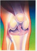
As mentioned previously the Popliteus is a muscle used mostly for kinesthetic awareness and stability in the knee. When an athlete pivots quickly or changes direction the Popliteus is put to work. When observing the picture below you can see how the muscle acts as a connection between the medial and lateral knee structures.


This means as the medial compartment experiences tension and the lateral side experiences compression the Popliteus receives this sensory feedback and stabilizes the knee. The opposite is also the same when the lateral side experiences tension and Medial experiences compression. This type of interaction occurs in the knee constently as an athlete brings the knee from flexion to extension and when performing pivoting moves.


So we need to rehabilitate it in a way that allows it to learn this function. However, how do you teach this function without putting stresses on the knee in an injured patient?

Exercises for the Popliteus

Exercises should consist of non-weight bearing and weight bearing exercises.

1. Using a resistant band attach one end to a stable service and the other around the forefoot of the affected leg. Using a wall for support begin the exercise with the affected leg unsupported at the patients side. From the start position begin flexing the knee and bringing the foot behind the stance leg as if attempting to touch your toe to your opposite hip. This creates flexion of the knee and internal rotation of the tibia. This motion will contract and activate the Popliteus. Now return to the starting position slowly to also train the Popliteus to contract eccentrically as well.


2. Stepping task - This exercise is used for progression and performed to challenge the Popliteus in a more functional manner. Using a small step up place one foot on the step allowing the knee to be maintained in a slightly flexed position. With the opposite leg off of the step bring yourself to a fencer type of stance.


Now while maintaining the supported knee in flexion begin different stepping manouvers. First going to 3 oclock back to start, 12 oclock and then 9 oclock. This exercise trains the Popliteus on how to prepare for cutting motions which will involve a forceful planting of the foot and manoeuvring in different directions.

The exercise can be further challenged by using verbal or visual cues to create a spongtanoise stepping action as apposed to the patient knowing where they will be going. Tossing a ball or using a racket can also make things more sports specific depending on the athlete. In addition, one can challenge the exercise by adding a blindfold to remove visual stimulus.

Once a patient displays proper technique in this exercise they may now progress to the same exercise above but using an unstable service.

3. Stepping Task Unstable Surface

4. The last exercise is done to train the Popliteus in a manner that challenges it to receive tension and then create a reflexive contraction. This involves a series of jumping task using pylons.

Standing alongside a pylon with one foot supported the patient jumps to the opposite side. The patient now has three options upon landing.

a) Land on the opposite leg they took off from allowing the Popliteus to be trained concentrically
b) Land on the same leg they took off from allowing the Popliteus to be used in a concentric to eccentric manner
c) Do option b followed by quickly jumping to the opposite side of the pylons allowing the Popliteus to be trained concentrically to eccentrically and then quickly concentrically again. This emulates a very quick stopping task or explosive take off manouver which man occur in multiple sports.


In summary, these are just a few quick exercises that can used in preseason training for athletes or any patient with knee problems. Dr. Wayne Button, BSc, DC


Nyland, J. (2005). Anatomy, Function, and Rehabilitation of the Popliteus Musculotendinous Complex Journal of Orthopaedic and Sports Physical Therapy DOI: 10.2519/jospt.2005.1414

Saturday, January 9, 2010

The Popliteus Muscle – Why does the back of my knee hurt?

The knee is one of the most complex joints in the human body. It is impossible to summarize such a topic with the understanding of only one muscle. However, certain muscles in the human body appear to be more important then others. With regards to the knee the Popliteus is one of these muscles. In this column I will attempt to explain the importance of the Popliteus Muscle in an injured knee and the rehabilitative process.


Anyone can open an anatomy textbook and retrieve the basic information about a muscle so how about I save you some time?


Popliteus Muscle:


O – Lateral Surface of the lateral condyle of the femur
Attachment to the lateral meniscus
I – Upper part of the posterior surface of the tibia
A – Medial rotates the tibia
Inn – Tibial Nerve (Moore, 2006)


Basic anatomy has its importance but to truly understand injuries one has to grasp the concept of muscles working in synergy together. Sometimes this ideology is referred to as the Kinetic Chain.


Some of the most important anatomical aspects of the Popliteus muscle will not be found in your basic textbook but in research. The Popliteus muscle is one of the most researched muscles in the knee and is always being debated upon its main role. Here are some important anatomical concepts you should know.


1. The Popliteus muscle plays an important role in “unlocking” the knee joint. When flexion and extension occurs the Popliteus is involved in rotating the Tibia and Femur. This action allows the two bones to deviate from one another and now the bones will not “collide” or “lock”. This mechanism leads to a smooth movement of flexion and extension throughout the knee.


This gives good indication to the importance of soft tissue therapy to the Popliteus muscle. When patients present with injuries that don’t allow a smooth or full range of motion the Popliteus muscle could be the culprit. With a knee that is “catching” the Popliteus muscle should be “released”. Releasing the Popliteus muscle can be done with heat, stretching, Myofascial Release Therapy, Active Release Technique or Graston Technique. (Manual Therapy Techniques)


2. The Popliteus muscle is what we call a “feed forward” muscle. Other “Feed Forward” muscles include the Multifidus in the back and Supraspinatus muscle in the shoulder. These are muscles that can actually “turn on” or “fire” before movement even occurs amongst the joint. Furthermore, these muscles have a high amount of receptors making them very important for proprioception and balance training. This is important to know because any patient who has an “unstable” joint or a past knee dislocation can vastly benefit from a rehabilitative program which exercises the Popliteus muscle.


In addition, this knowledge is a good indication that athletes may benefit from a preseason exercise routine, which involves the Popliteus muscle. By training a muscle involved in proprioception this may lower the incidence of knee injuries. This same concept is already being explored with preseason prevention of ankle injuries and acl tears by certain workout regiments.


3. The Popliteus muscle also works in synergy with the posterior cruciate ligament (PCL). Anatomical studies have shown that by actually cutting the Popliteus muscle tension increased in the PCL. The same is true otherwise that by increasing the force through the Popliteus, tension has been lowered in the PCL. This concept makes sense when you observe the following picture. When you look at the Popliteus and PCL together they almost both make a hammack for the knee joint allowing the bones to rest on their surfaces. It makes sense that if you were to injury, tear or have one weak the other would need to support the load.


This details the fact that by maybe strengthening the Popliteus muscle PCL injuries would decrease. Furthermore, if you have a weak and dysfunctional Popliteus you may develop PCL insufficiency or obtain an injury in the future.


Need more details? The Popliteus also:
- helps in movement of the lateral meniscus
-Is involved in posteriorlateral stability of the knee
-Plays an important component in down hill running
-Has attachments to all of the following


–Distal MCL
–Fibular Head
–Lateral Meniscus
–PCL
–Femur
–Tibia


These are just a few concepts of why I believe the Popliteus is thee most important muscle of the knee. Even if the muscle itself is not injured addressing it may give significant results. The old concept of only treating what is injured is dead and understanding the body as a whole is starting to evolve.


So how do I exercise/rehabilitate the Popliteus muscle?
Tune in for a rehab protocol of the Popliteus next blog


Nyland, J. (2005). Anatomy, Function, and Rehabilitation of the Popliteus Musculotendinous Complex Journal of Orthopaedic and Sports Physical Therapy DOI: 10.2519/jospt.2005.1414

Monday, December 21, 2009

Merry Christmas


Hey guys just a quick note wishing everyone a Merry Christmas and a Happy New Year. The blog will be taking a small break for the next two weeks. I don't know if i'll be posting anything over Christmas because of lack of time but if so keep checking in.

These last few months have been great as the start of my new blog. What originally was a pass time to keep me up to date on research and current has lead to over 2000 viewers a month. I expect great things for 2010 and have a list of future blogs. Any ideas or questions please feel free to email me and ill work on those blogs for 2010.

Thank you for all the patients, friends, viewers and other professionals which have made comments on the blog. Everyone have a great Christmas. Dr. Wayne Button, BSc, D.C


Sunday, December 13, 2009

Patellofemoral Pain Syndrome- Where exactly is the pain coming from?

The knee of an athlete is often a diagnostic dilemma. The knee obtains important biomechanical function and is supported by an array of ligaments and muscles. For these reasons it can be difficult to pinpoint the specific structure which is injured. Such a concept is to be blamed for the creation of Patellofemoral Pain Syndrome (PFPS),


What exactly is PFPS?

When you think about it what exactly does this diagnosis tell you? Essentially all that this means is we know there is pain and we know it has something to do with the Patella-Femoral Joint. In my opinion this is not a real diagnosis. What if we made clinical conclusions like this for everything? Chronic Neck Pain Syndrome or Sore Elbow Disease?

In reality, PFPS is an umbrella term to describe any possible causes of pain which can be attributed to the Patella-Femoral Joint. Such as:

-Chondromalacia Patella - Softening of the cartilage under the patella
-PatellaFemoral tracking problems
-Infrapatellar Fat Pad being impinged (Hoffa's Disease)


 This is why I often tell colleagues and those which attend my seminars to not worry about the diagnosis. What you need to find out is WHAT EXACTLY IS CAUSING THIS PAIN?

Originally many used to take a structural approach to PFPS. Measurements of one's Q angle or a patient's knee valgus was observed and often chondromalacia patella was diagnosed. However, if structural abnormalities were deemed the cause how does one explain the benefits from conservative treatment? The patients Q angle has not changed. Furthermore, not everyone with a high Q angle has knee problems?


Researchers are starting to progress away from the structural approach of PFPS and asking what is actually causing the pain?

Ok so what are my options?

A biomechanical fault, muscle imbalance or pathophysiological process which leads to:

1.Inflammation of the synovial lining
2.Inflammation of the fat pad
3.Increase in intra articular pressure

4.Hyperinnervated Lateral Retinaculum  

To fully understand you have to address the signs and symptoms

PFPS can be exaggerated with running and climbing up and down stairs. This mechanism of injury is simply due to overuse or excessive force. Such activities as climbing stairs can produce forces 3 times one's body weight. A loss of Tissue Homeostasis occurs and now the joints synovial lining becomes inflamed (#1). This concept is a pretty easy one to understand.

However, how does one explain the movie sign (Pain in the knee with a prolonged period of sitting)? One does not put excessive force through the knee as they sit. There are two leading theories for the movie sign. While sitting we may predispose our knee to change normal position and now structures my impinge a once swollen inflamed synovial lining (#1). The second theory is that as we sit our knee's intra articular pressure increases and thus creates pain (#3). Studies have shown that the knee is an extremely fibrotic and capsular entity. When put in prolonged flexion we may impede venous outflow and increase arterial blood flow to and from the knee. This change in blood flow can increase the pressure within our knee as we sit.

Reasoning for these causes of pain is the popular treatment called McConnell taping. This is a taping method which has shown to relieve pain. However, the results are not always consistent and many wonder why. It can be purposed that by taping the patella in a more neutral position we are preventing it from impinging structures and thus resolving the patient's pain (#1, #2).

Lastly a maltracking patella has also been associated with PFPS. What studies have observed is anatomically those who suffer from PFPS have a hyperinnervated lateral retinaculum. Furthermore, these subjects presented with more nociceptive fibers within the nerves. The theory is the patella tracking medially (usually) will produce tension along the lat retinaculum. Now the nerves are put under traction and start to experience myxoid degeneration. In an attempt to heal itself the body now produces more neural drive and stimulation to that area thus creating a hyperinnervated structure and pain is produced (#4).

Still Don't Believe Me?

Check out the article posted below by Scott F Dye. This is one of the most intriguing and interesting articles I have ever read. Scott which is an orthopedic surgeon (I believe?) has actually been diagnosed with chondromalacia patella in the past. He attempts to cut himself open and inspect the cause of his knee pain. When probing his patella cartilage no pain is felt. However, pain is accentuated when probing his synovial lining and Hoffa fat pad. Furthermore injecting himself with saline increases his intra-articular pressure and produced extreme pain. This article has given valuable premise to support 3/4 theories of what causes PFPS. Imagine... the things we do for science (Left probing cartilage, Right probing synovial lining).


In summary, as professionals we need to steer away from umbrella terms such as PFPS. As a student I found this diagnosis extremely confusing and more so difficult to explain to patients. It wasn't until exploring the mechanism of pain in which I become more inclined to make such a diagnosis. To create consistency in research and amongst colleagues we need to address the actual form of PFPS were discussing. In addition, exploring this notion lays great premise on how we can guide our treatment. This is one condition where the diagnosis may actually be made after treatment is initiated. Dr. Wayne Button, BSc, D.C

Dye, S., & Vaupel, G. (1994). The Pathophysiology of Patellofemoral Pain Sports Medicine and Arthroscopy Review, 2 (3), 203-210 DOI: 10.1097/00132585-199400230-00004

Sanchis-Alfonso V, & Roselló-Sastre E (2000). Immunohistochemical analysis for neural markers of the lateral retinaculum in patients with isolated symptomatic patellofemoral malalignment. A neuroanatomic basis for anterior knee pain in the active young patient. The American journal of sports medicine, 28 (5), 725-31 PMID: 11032232

Naslund, J., Walden, M., & Lindberg, L. (2007). Decreased Pulsatile Blood Flow in the Patella in Patellofemoral Pain Syndrome The American Journal of Sports Medicine, 35 (10), 1668-1673 DOI: 10.1177/0363546507303115


-

Saturday, December 12, 2009

Low back pain while running..a real pain in the but. Part 2:

Refer to past column Low Back Pain while running..a real pain in the but. Part 1:

Inspired by my own past experience. 

So how do I fix this?

To help fix this problem you have to train yourself to achieve an efficient running style. You need to combine loosening your tight Psoas and activating your inhibited Glut Max.

1. The tight Psoas - There are multiple ways to loosen up a muscle before activity. People can use heat, massage, different soft tissue therapy techniques and stretching. Opinions on stretching differ significantly. Some say it''s beneficial and some say it isn't. The truth of the matter is there is no conclusive answer. However, if a muscle is relatively tight or leading to pain I recommend doing specific stretches before a run.

Stretches for the Psoas:


2. The inhibited Glut Max - Ever watch elite runners before a race. Often you can see runners pounding their fist against their Gluts. That's because their trying to wake their Glut Max up before attempting to use it. I know it seems weird but before your next run try hitting your Gluts with your fist to wake them up. This action can stimulate proprioceptive and biofeedback to the muscle.

One should also try incorporating basic Glut Max exercises into their training program as well. If too many of these exercises are done before running it may actually fatigue the Glut Max and exaggerate the problem. However, just a few simple repetitions to stimulate your Gluts should suffice.

Glut Max Exercises


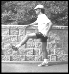
3. Putting it all together - Before running try to emulate your running technique when warming up. Against a wall for balance, just try swinging your leg or performing kick backs as if you are running. You want to teach your back to stay neutral as all the movement is carried by your hips. Doing this before hand also allows you to gauge the flexibility in your hips and awareness of your Glut. If still not suffix try a few more stretches or exercises. For training purposes this can also be done with a resistance cord or ankle weights.


In summary, a few simple stretches and exercises before a run or race can help a lot. If your problem persists get it assessed by a trained professional. Dr. Wayne Button, BSc, D.C

Liebenson, C. (2009). Functional training of the gluteal muscles☆ Journal of Bodywork and Movement Therapies, 13 (2), 202-204 DOI: 10.1016/j.jbmt.2009.01.004

Wednesday, December 9, 2009

Low back pain while running..a real problem in the but? Part 1

There are a number of causes of low back pain in recreational joggers. However, what many people may not know is often the pain can be attributed to other areas rather than the low back.


To believe the body works as different compartments is a false notion. Our body works as a complete unit and in synergy together during exercise. Jogging and running is no exception to this concept. This is why back pain usually can be caused by problems in your hips and buttock region. That's right ...your low back pain could be significantly reduced by addressing muscles in your but.

I don't understand??

1. Your Gluteus Maximus is the biggest muscle in your buttocks area. Although this muscle has many roles in running it is involved in extending your leg through the swinging phase. When the muscle is not firing properly or inhibited, your leg will not extend to a full range of motion it is capable of. Inhibited simply is a term used to describe a muscle which is weak not due to pain or neurological cause.


When the Gluteus Maximus is not as strong or efficient the body has to compensate by finding other ways to achieve the range of motion it would like. So what runners will do without even realizing it is extend or hike their back to gain that extra range of motion.


2. A second muscle involved in this type of faulty running behaviour is the Psoas muscle. This is a muscle which crosses the front of the hip joint. While running the Psoas is involved in flexing the hip forward. Needless to say the Gluteus Maximus and Psoas are contently battling against one another as you run. One helps bring your leg forward as the other brings it back..over and over again.


This is why a Psoas muscle which is tight can also decrease the extension you achieve in your hips. Thus causing you to compensate again by arching your back.

3. Putting it all together - This combination of a weak Glut Max and a tight Psoas muscle will lead a runner towards a compensating gait. Now your back is exposed to extra forces it does not need on a consistent basis.

Its important when running to allow most of the movement to occur from your hips and allow the back to stay neutral. By creating this running technique back pain may be prevented and decreased significantly.

How do I know I have this problem?

Well any type of back pain should be addressed by a trained professional. This is because there can be multiple causes for the pain. It is important to rule out a serious pathology. However, there are ways to see if you have this particular problem. You may need a partner or again someone trained to observe different movement patterns.

1) Your gait - Sometimes this subtle hiking of your back can be seen from a partner while watching you run from a side view. Videotaping patients while running on a treadmill is a great learning tool to use as well.

2) The tight Psoas - You may just feel the tightness in general but can still use different test to see if it is tight.

On a gym bench or table lye on your back with your buttock at the end of the table. Allow your legs to hang off the bench freely. With your hands take one leg and bring your knee towards the chest. If the opposite leg comes off the table almost immediately or in an excessive range this can indicate a tight Psoas. This same test is often used by manual therapist and is called the Modified Thomas test.


3) A inhibited Glut Max- This is something that is very tricky to test for. That is because it is hard to assess how the Glut will function in isolation from other muscles. Seeing how much you can squat is not exactly a reliable measure. It's important to assess how does the Glut Max work in motion which resembles a running gait. Many manual therapist use what is called the Janda Prone Hip Extension Test (named after the famous neurologist Vladimir Janda).

This test should be assessed by someone who is trained to analyze movement patterns. Start by lying on your stomach upon a bench or table. While maintaining this position slowly extend one of your legs up towards the ceiling just a few inches off the table. A faulty movement pattern will be either a bent knee, hinging of the low back or a delayed response from your Gluteus Maximus. A normal test should show a smooth pattern of leg extension with the Glut Max doing most the work. When the back hinges or the knee is bent this is signs of other muscles helping the Glut Max or worse doing its job (Top Right & Lower Left faulty patterns)


In summary, back pain and running injuries can be a complicated manner. The answers are not always this easy and should be addressed by someone who is trained in such a topic.


How do I solve this problem? Stay tuned for Part 2.


LIEBENSON, C. (2007). Hip dysfunction and back pain Journal of Bodywork and Movement Therapies, 11 (2), 111-115 DOI: 10.1016/j.jbmt.2007.01.005

Sunday, December 6, 2009

I sprained my ankle months ago and it still hurts?


A sprained ankle is the most common sports injury. However, it usually is easy to diagnose and will eventually always heal. Sprained ankles are expected to be back to 90% recovery within 6 weeks (This may vary depending on the degree of sprain and other associated factors). However, what if it doesn't get better? What if your symptoms have not resided by that time? Believe it or not, a simple sprained ankle can often lead to long term problems.


1) Osteochondral Lesions/Osteocondritis Dissecans of the Talus

Often when an ankle is sprained there can be a sudden loss of stability. This can cause bones within the ankle to impact or collapse into one another causing an osteochondral lesion to form. An osteocondral lesion is when the cartilage surrounding a bone is torn, crushed and in more rare causes forms a cyst. Cartilage is the cushioning our bones and joints are surrounded by to help with impact forces. With the cartilage now torn or disrupted it may break off with a small piece of bone and be wedged or displaced inside a joint.


Symptoms often include persistent pain with activity. The pain may be deep within the joint and can occur long after the pain felt from a sprained ankle has resided. The ankle may also experience catching, snapping or locking which it hasn't experienced before. There have been reports of asymptomatic cases and individuals healing spontaneously, but this is often amongst a younger population. Furthermore, these lesions can at times progress to create osteoarthritis within the ankle joint.

Do not assume you do not have this injury because you had radiographs taken. The lesions are small (2-5mm) and flake like. Furthermore, special views are commonly used to spot the injury which will often not be included in a normal series of ankle radiographs. Observe below (A & B no lesions is shown but can be seen on view C). Needless to say this injury is difficult to spot. If easily accessible an MRI should be taken as it has shown optimal diagnostic value.


Osteocondral lesion can be treated conservatively and surgically. However, there has been a lack of research to determine what factors play into the decision making process. Most children or adolescents will resolve the lesion because of their healing properties but adults often carry out the option of surgery.

2)Complex Regional Pain Syndrome/ Reflex Sympathetic Dystrophy

There is no standard definition for CRPS, as nobody has determined what causes the condition. Essentially the syndrome can be defined by its simplistic name "Complex Regional Pain". The syndrome involves a complex array of symptoms in a particular region which leads to pain. However, CRPS has a neurological component explaining the symptoms.

The cause is often attributed to the neurological system becoming confused. When someone experiences an injury the bodies nervous system reacts by producing pain, increasing swelling, changing color due to inflammation ect. However, with CRPS the nerves now are hyper-vigilant, meaning the littlest touch or activity can set of a chain reaction replicating a more serious injury. Reports have shown incidents as small as a paper-cut to cause CRPS. Simple events such as walking or putting on your socks can be interpreted by the ankles nervous system as getting hit by a hammer.


Symptoms will include pain disproportional to the event causing the symptoms originally. Light touch or the application of any form of heat or coldness may be taken as intolerable. Nerves which innervate blood vessels and sweat glands are also affected. Symptoms of this nature may will appear as excessive swelling by the ankle, changes in color, changes to the appearance of the toe nails, hair loss and even sweating when not indicated. In Addition, muscles around the ankle with time may become weakened and get smaller in appearance.

Diagnosing CRPS isn't an easy task. Radiographs, MRI and common physical examination procedures can give a good implication of a diagnosis but not always confirm. Often special imaging is carried out to assess the bodies thermo graphic patterns.

3) Ankle Impingement Syndrome

The body works on a simple concept that when a structure gets damaged it will try to repair itself. Furthermore, whenever a repetitive force or even a constant pressure is applied to a structure it will react by growing, shaping or forming a new position to compensate for this pressure. This concept is how braces work for shaping ones teeth properly.

In the ankle there are a lot of ligaments, muscles and joints. Often when repetitive trauma or strain is applied to one of these structures it will compensate similar to what was described above. Eventually the small bit of cartilage surrounding the bones or ligaments will be pulled on or experience "traction". This constant repetitive action will lead to the formation of little bone and cartilage spurs. These spurs will also occur after repetitive ankle sprains. This process is called osteophytosis.


The spurs formed can often not present any problem at all. However, occasionally bone spurs (also called "osteophytes") will "impinge" structures such as ligaments, tendons and scar tissue from past injuries.

Signs and symptoms may include an ankle which feels unstable or weak. Tenderness or a "pinching sensation" may occur when the foot is brought through flexion and extension. A painful clicking may also be observed during certain movements of the ankle. Impingement can occur in the front and back of the ankle. Usually squatting or bringing your toes towards your head will be painful in anterior impingement. Bringing your toes downward or rising on your toes will produce pain in posterior impingement.


5) Cuboid Syndrome

The cuboid is a small bone that lies on the outside of the foot. Cuboid syndrome is when this bone experiences excessive movement or a collapsing effect. The excessive movement can cause irritation of the surrounding joint capsule and ligaments or put strain on a particular muscle which attaches to the cuboid called the fibularis longus.

Up to 4% of athletes can experience cuboid syndrome with a particular high rate in ballet dancers. However, up to 6.7% of those experiencing ankle sprains can develop cuboid syndrome. When the foot is forced into this position the fibularis longus muscle experiences tension thus jamming the cuboid bone and forcing it to shift out of place.

Patients with cuboid syndrome may present with pain directly over the cuboid and radiating down the outside of the foot. Pain is often associated with a weight bearing position but can occur otherwise. Pain can be experienced with squatting or on toe off during a running gait. Weakness can often be a secondary complaint.

The good news is cuboid syndrome has been shown to be successfully treated with conservative treatment including rehab, manipulation, taping and the placement of a small felt pad under the cuboid preventing it from collapsing.


In summary, if your experiencing new symptoms after an ankle sprain get it assessed by a trained professional. The complaint may be something completely new but sprained ankles often lead to problems down the road. Assess the problem early, listen to your clinician and take precautions on your first ankle sprain to reassure these problems don't persist.

Other injuries that may occur after a sprained ankle or be confused with previous mentioned conditions are:

-Os trigonum
-Fibularis Longus Subluxation
-Sinus Tarsi Syndrome
-Osteoarthritis
-Nerve Palsy

References


-


Choi WJ, Park KK, Kim BS, & Lee JW (2009). Osteochondral lesion of the talus: is there a critical defect size for poor outcome? The American journal of sports medicine, 37 (10), 1974-80 PMID: 19654429

PONTELL, D. (2008). A Clinical Approach to Complex Regional Pain Syndrome Clinics in Podiatric Medicine and Surgery, 25 (3), 361-380 DOI: 10.1016/j.cpm.2008.02.011

Jennings, J. (2005). Treatment of Cuboid Syndrome Secondary to Lateral Ankle Sprains: A Case Series Journal of Orthopaedic and Sports Physical Therapy DOI: 10.2519/jospt.2005.1596